40 diagram of the brain without labels
Brain: Atlas of human anatomy with MRI - e-Anatomy The module on the anatomy of the brain based on MRI with axial slices was redesigned, having received multiple requests from users for coronal and sagittal slices. The elaboration of this new module, its labeling of more than 524 structures on 379 MRI images in three different views and on 26 anatomical diagrams, took more than 6 months. Lobes of the brain - Queensland Brain Institute ... The brain's cerebral cortex is the outermost layer that gives the brain its characteristic wrinkly appearance. The cerebral cortex is divided lengthways into two cerebral hemispheres connected by the corpus callosum.Traditionally, each of the hemispheres has been divided into four lobes: frontal, parietal, temporal and occipital. (Wikimedia)
Neuron Diagram Unlabeled neuron, (1). axon, cell body, dendrites, nucleus, terminal. Unlabeled diagram of a motor neuron (try labeling: axon, dendrite, cell body, myelin, nodes of Ranvier, motor end plate).Read the definitions, then label the neuron diagram below. axon - the long extension of a neuron that carries nerve impulses away from the body of the cell.

Diagram of the brain without labels
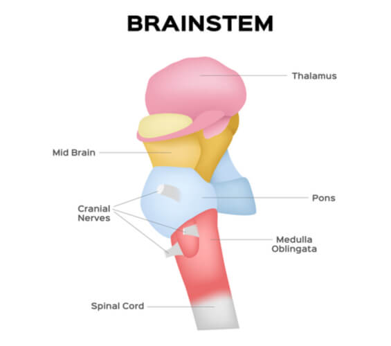
Brain: Anatomy, Pictures, Functions, and Conditions The Brain Stem. PIXOLOGICSTUDIO/SCIENCE PHOTO LIBRARY / Getty Images. The brainstem is an area located at the base of the brain that contains structures vital for involuntary functions such as the heartbeat and breathing. The brain stem is comprised of the midbrain, pons, and medulla. 3. Diagram of the Brain and its Functions - Bodytomy Given below is a labeled diagram showing the brain stem and its related structures. Brain Stem and Structures Cerebellum The word 'cerebellum' literally means little brain. It is the second largest part of the brain, and is located at the back, below the occipital lobe, beneath the cerebrum and behind the brain stem. Parts of the brain: Learn with diagrams and quizzes | Kenhub Labeled brain diagram First up, have a look at the labeled brain structures on the image below. Try to memorize the name and location of each structure, then proceed to test yourself with the blank brain diagram provided below. Labeled diagram showing the main parts of the brain Blank brain diagram (free download!)
Diagram of the brain without labels. Anatomy of the Brain: Structures and Their Function The midbrain or mesencephalon, is the portion of the brainstem that connects the hindbrain and the forebrain. This region of the brain is involved in auditory and visual responses as well as motor function. The hindbrain extends from the spinal cord and is composed of the metencephalon and myelencephalon. Mapping the Brain - BrainFacts Mapping the Brain. The cerebrum, the largest part of the human brain, is associated with higher order functioning, including the control of voluntary behavior. Thinking, perceiving, planning, and understanding language all lie within the cerebrum's control. The top image shows the four main sections of the cerebral cortex: the frontal lobe ... Human Brain - Structure, Diagram, Parts Of Human Brain The brain diagram given below highlights the different lobes of the human brain. Where is the Brain located? The brain is enclosed within the skull, which provides frontal, lateral and dorsal protection. The skull consists of 22 bones, 14 of which form the facial bones and the remaining 8 form the cranial bones. Brain Label - The Biology Corner Image of the brain showing its major features for students to practice labeling. Answers are included.
4,689 Human Body Anatomy With Labels Stock Photos and ... human body anatomy with labels Stock Photos and Images. 4,689 matches. Page of 47. The human digestive system, digestive tract or alimentary canal with labels. Labelled with UK spellings and labels like those in the GCSE syllabus. Human digestive system, digestive tract or alimentary canal including text labels. 14 Informative Facts, Diagram & Parts Of Human Brain For Kids The brain weighs just about two to three pounds and appears like a walnut. The brain is comprised of three main regions — cerebrum, cerebellum, and brainstem (3). Let us discuss these parts and their functions in more detail (1) (3) (4). Cerebrum: The cerebrum is the largest part of the brain. Nervous System - Label the Brain Nervous System - Label the Brain Nervous System - Brain Name: Choose the correct names for the parts of the brain. ( 1) (2) (3) (4) (5) (6) (7) (8) ( 9) This brain part controls thinking. (10) This brain part controls balance, movement, and coordination. (11) This brain part controls involuntary actions such as breathing, heartbeats, and digestion. Human Nervous System - Diagram - How It Works | Live Science It consists of the brain, spinal cord and the retinas of the eyes. The Peripheral Nervous System consists of sensory neurons, ganglia (clusters of neurons) and nerves that connect the central...
Brain (Human Anatomy): Picture, Function, Parts ... • The cortex is the outermost layer of brain cells. Thinking and voluntary movements begin in the cortex. • The brain stem is between the spinal cord and the rest of the brain. Basic functions like... Anatomical diagrams of the brain - e-Anatomy These original illustrations and diagrams of the brain were created from 3D medical imaging reconstructions and then redrawn and colored using Adobe Illustrator. These anatomical charts include the main diagrams necessary for medical students, nursing students, residents, practitioners, anatomists to study the anatomy of the brain, to ... The Human Brain (Diagram) (Worksheet) - Therapist Aid The Human Brain Diagram is a versatile tool for psychoeducation. The diagram separates the brain into six major parts, and provides a brief description of the functions carried out by each section. Discussion of the brain, and how it works, can be a powerful way to explore many topics. The Human Brain - Visible Body The brain gives us self-awareness and the ability to speak and move in the world. Its four major regions make this possible: The cerebrum, with its cerebral cortex, gives us conscious control of our actions. The diencephalon mediates sensations, manages emotions, and commands whole internal systems. The cerebellum adjusts body movements, speech ...
Labeled Diagrams of the Human Brain You'll Want to Copy ... The average dimension of the adult human brain is 5.5 inches in width and 6.5 inches in length. The height of the human brain is about 3.6 inches and it weighs about 4 to 5 lbs at birth and 3 lbs in adults. The total surface area of the cerebral cortex is about 2,500 cm2 and when stretched, it will cover the area of a night table.
Human Brain Diagram - Labeled, Unlabled, and Blank Human Brain Diagram - Labeled, Unlabled, and Blank Click here to download a free human brain diagram. Learn the parts of the human brain with these convenient printables for students and teachers. Tim's Printables 36k followers More information Human Brain Diagram - Labeled, Unlabled, and Blank
Label Brain Diagram Printout - EnchantedLearning.com Label the Brain Anatomy Diagram The Brain Read the definitions below, then label the brain anatomy diagram. Cerebellum - the part of the brain below the back of the cerebrum. It regulates balance, posture, movement, and muscle coordination. Corpus Callosum - a large bundle of nerve fibers that connect the left and right cerebral hemispheres.
Brain - Human Brain Diagrams and Detailed Information Brain cells can be broken into two groups: neurons and neuroglia. Neurons, or nerve cells, are the cells that perform all of the communication and processing within the brain. Sensory neurons entering the brain from the peripheral nervous system deliver information about the condition of the body and its surroundings.
A Neurosurgeon's Overview the Brain's Anatomy The cerebrum or brain can be divided into pairs of frontal, temporal, parietal and occipital lobes. Each hemisphere has a frontal, temporal, parietal and occipital lobe. Each lobe may be divided, once again, into areas that serve very specific functions.

In this diagram it shows the different parts of the brain, as well as the functions they perform ...
Brain Chart Maker - 100+ stunning chart types — Vizzlo A brain chart is a fun way to visualize the things that are on your mind. A great tool to depict the functions and anatomy of the brain - ideal for school presentations. How to make a brain chart with Vizzlo? Click on the "DATA" tab to add, remove or edit records. You can also adjust the size of each area by dragging the slider.
Label The Brain - Mr. Barth's Class You won't label the parts of the brain on this website, but you'll familiarize yourself with the location of the parts and their basic functions. Lobes of the Brain Click on the link to the left to label the lobes of the brain. See how quickly you can do it with 100% accuracy. Lobes and Neuron Diagram
Simple diagram of brain - Healthiack Best viewed on 1280 x 768 px resolution in any modern browser. Simple diagram of brain 1352. Simple diagram of brain 1360. Simple diagram of brain 1368. Simple diagram of brain 1376. Simple diagram of brain 1378. Simple diagram of brain 1384. Simple diagram of brain 1388. Simple diagram of brain 1393.
Diagram Of Brain with their Labelings and Detailed Explanation The midbrain is the smallest region of the brain, found at the centre of the brain, between cerebral cortex and hindbrain. It comprises tectum, cerebral peduncle, tegmentum, cerebral aqueduct, substantia nigra, several nuclei and fasciculi. The midbrain is responsible for hearing, vision, sleep cycle, temperature regulation, alertness, etc.
Brain Label #apbiology #ap #biology #human #body | Human ... Neurons are the brain's rock stars. But without the glial cells — astrocytes, microglia and oligodendrocytes — there would be no show at all. A diagram by Arne Hurty for Stanford Medicine Magazine Fall 2009.
Parts of the brain: Learn with diagrams and quizzes | Kenhub Labeled brain diagram First up, have a look at the labeled brain structures on the image below. Try to memorize the name and location of each structure, then proceed to test yourself with the blank brain diagram provided below. Labeled diagram showing the main parts of the brain Blank brain diagram (free download!)
Diagram of the Brain and its Functions - Bodytomy Given below is a labeled diagram showing the brain stem and its related structures. Brain Stem and Structures Cerebellum The word 'cerebellum' literally means little brain. It is the second largest part of the brain, and is located at the back, below the occipital lobe, beneath the cerebrum and behind the brain stem.
Brain: Anatomy, Pictures, Functions, and Conditions The Brain Stem. PIXOLOGICSTUDIO/SCIENCE PHOTO LIBRARY / Getty Images. The brainstem is an area located at the base of the brain that contains structures vital for involuntary functions such as the heartbeat and breathing. The brain stem is comprised of the midbrain, pons, and medulla. 3.


Post a Comment for "40 diagram of the brain without labels"